Facility


Basic Information
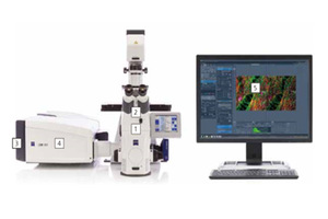
Vendor:OXFORD INSTRUMENTS
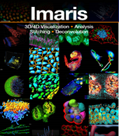
Instrument:Bitplane Imaris 9.3.1
Reservation Call: (021)31246791
Location: Room A101-3, Building of Life Sciences
Instrument Specification
Reconstruct and Visualize complex 3/4D microscopy datasets up to terabytes in size.
Precisely detect and segment objects using the wizard-driven segmentation tools in Imaris.
Calculate size and shape parameters, signal intensity, and relative position in space for all detected objects.
Track objects over time (4D tracking) using the highly reliable algorithms, and display motion related parameters: speed, displacement vectors, track straightness, cell divisions, lineage tree, etc.
Detect, trace and measure neurons’ dendritic trees, axons, spines, blood vessels and microtubules.
Detect cells based on cytoplasm labeling or based on plasma membrane labeling, and examine relationships between cells and cellular components.
Check colocalization of fluorescent signal from various components in 3D over time.
GPU-accelerated Deconvolution to improve image quality.

Basic Information
Vendor:PE
Instrument: Opera Phenix
Reservation Call: (021)31246791
Location : Room A101-3, Building of Life Sciences
Instrument Specification
Spinning-disk optics and careful synchronization of laser excitation and camera exposure minimize bleaching and phototoxicity.
Proprietary automated water-immersion objectives with very high numerical aperture deliver and capture more photons and provide higher resolution in XYZ than conventional air objectives.
Up to two large-format sCMOS cameras deliver low noise, wide dynamic range, and high resolution – perfect for sensitive and quantitative measurements at short exposure times.
The Opera Phenix system's Synchrony Optics feature a microlens-enhanced Nipkow disk with increased transmission rates for fast, sensitive, true-multipoint confocal imaging. The system's unique dual-view layout separates the excitation and emission paths for fluorophores with overlapping spectra by creating non-overlapping pinhole patterns in the sample. This minimizes crosstalk during simultaneous confocal acquisition by 98% on average, so you can simultaneously acquire images of the nucleus (labeled with Hoechst) and of the cytoplasm (labeled with GFP, for example) and up to two more markers – without limiting sensitivity.

Basic Information
Vendor: NIKON
Instrument: A1&SIM&STORM
Reservation Call: (021)31246791
Location : Room A101-5, Building of Life Sciences
Instrument Specification
Combined with laser scanning confocal (Confocal), structured illumination microscopy (SIM) and stochastic optical reconstruction microscopy (STORM) technology.
The spatial resolution of SIM is up to 20 nm, which can realize multi-channel 3D fluorescence imaging.
High-speed piezoelectric 3D scanning electric stage, 32-channel spectral imaging, perfect focus system (PFS) are suitable for long-term observation of living cells.
Suitable for immune calibration of fixed cells and fluorescent living cells.
Function Application
Cell migration and cytoskeleton; cell division and cell cycle; cell signal transduction; tissue differentiation and development; vesicles and protein transport; physiology and neuroscience; calcium signal research; protein-DNA interaction; Host - pathogen interaction; Cancer research; Pharmacology research; Biophysics research. Research on living microorganisms, proteins, viruses, subcellular structures, etc.
Basic Information
Vendor:ZEISS
Instrument:LSM 880 with Airyscan
Reservation Call: (021)31246791
Location : Room A101-4, Building of Life Sciences
Instrument Specification
Airyscan detector for 2x increase in spatial resolution in each dimension (120 nm lateral and 350 nm axial resolution), 4-8 increase in SNR.
19 fps @ 512x512 at Fast Airyscan mode.
Spectra imaging and Linear unmixing with a 32 channels spectral detector for 6-9 color fluorescent imaging.
Advance confocal function, such as FRAP, FRET, TILE, Multi-dimension with Experiment Designer Module.
Function Application
Cell Biology Imaging
Molecular Localization
Pharmacokinetics

复旦大学 代谢与整合生物学研究院 版权所有
Copyright@2018fudan.edu.cn All rights reserved.
上海市杨浦区淞沪路2005号。邮编:200438。电话:021-31242078,021-31242079
 Fudan Email
 Fudan Ehall